Article published on a pioneering X-ray technique unveiling the 3D nanoscale architecture of functional materials
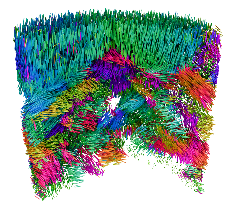
In a recent publication, we describe the development of a pioneering X-ray technique to probe the 3D orientation of a material’s building blocks at the nanoscale. The technique allows the visualization of crystal grains, grain boundaries and defects - critical factors that govern material performance.

Zoom into the micro or nanostructure of functional materials, both natural and manmade, and you’ll find they consist of thousands upon thousands of coherent domains or grains - distinct regions where molecules and atoms are arranged in a repeating pattern. Such local ordering is inextricably linked to the material properties. The size, orientation, and distribution of grains can make the difference between a sturdy brick or a crumbling stone; it determines the ductility of metal, the efficiency of electron transfer in a semiconductor, or the thermal conductivity of ceramics. It is also an important feature of biological materials: collagen fibres, for example, are formed from a network of fibrils and their organisation determines the biomechanical performance of connective tissue. These domains are often tiny: tens of nanometres in size. And it is their arrangement in three-dimensions over extended volumes that is property-determining. Yet until now, techniques to probe the organisation of materials at the nanoscale have largely been confined to two-dimensions or are destructive in nature.
Using X-rays generated by the Swiss Light Source, we have together with our collaborators succeeded in creating an imaging technique to access this information in three-dimensions. This technique is known as X-ray linear dichroic orientation tomography, or XL-DOT for short. XL-DOT uses polarised X-rays from the Swiss Light Source, to probe how materials absorb X-rays differently depending on the orientation of structural domains inside. By changing the polarisation of the X-rays, while rotating the sample to capture images from different angles, the technique creates a three-dimensional map revealing the internal organisation of the material.
This work was carried out in collaboration with researchers from the Paul Scherrer Institute, ETH Zurich, the University of Oxford and the Max Plank Institute for Chemical Physics of Solids.
Original text by Miriam Arrell, Paul Scherrer Institute. The full version can be found external page here.
X-ray Linear Dichroic Tomography of Crystallographic and Topological Defects
A. Apseros, V. Scagnoli, M. Holler, M. Guizar-Sicairos, Z. Gao, C. Appel, L. J. Heyderman, C. Donnelly, J. Ihli
external page Nature, 636, 354–360 (2024)